Profiling a landscape of heterogeneous cell types and biomolecules has become imperative to develop precision medicine and address current research questions.

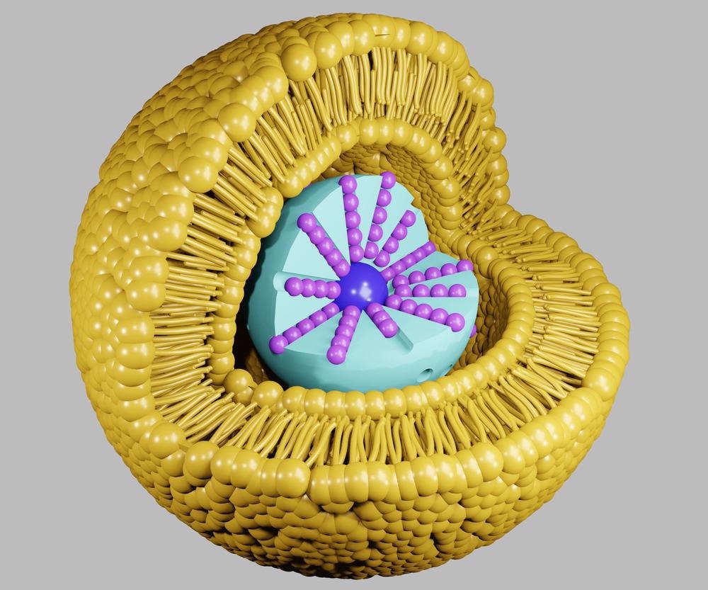
Study: Expanding the Multiplexing Capabilities of Raman Imaging to Reveal Highly Specific Molecular Expression and Enable Spatial Profiling. Image Credit: Love Employee/Shutterstock.com
In an article recently published in the journal ACS Nano, researchers presented a library of individually unique surface-enhanced Raman spectroscopy (SERS) nanoparticles (NPs), composed of approximately 60 nanometers gold (Au) core, a Raman reporter molecule, and a silica coating. They also demonstrated the application of prepared SERS NPs to target cultured cancer cells and profile human cancerous tissue sections.
SERS NPs in Raman Spectroscopy
The presence of metallic NPs or nanotextured metallic surfaces in SERS enhances the optical cross-section of Raman scattering. Modern hyperspectral imaging instrumentation combined with cutting-edge nanotechnology enables precise, fast, and efficient Raman analysis. The necessity for rapid profiling of disease tissues is burgeoning. Consequently, imaging technologies and high-throughput assays have gained considerable attention in the market. Research in fundamental biology to personalized treatment for patients requires biomarker assays to provide superimposed analyte signals that are segregated into their constituent components.
Molecular imaging of biological samples with SERS NP staining contrast agents leverages Raman spectroscopy to transmit the biomarker expression as multiplexed optical signals. Additionally, localized surface plasmon resonance (LSPR) of nanoscale gold uses nondestructive laser intensities to afford fast mapping speeds with high sensitivity. Thus, the Raman spectrum of SERS flavor serves as a reference to simultaneously discover and localize various protein targets.
Encapsulating consistent quantities of reporters in all NP batches is a difficult task. Multiplexing such NPs is challenging because measurements with multiple singly labeled NPs cannot be distinguished from those with NPs labeled with a combination of the same reporters. Additionally, at low concentrations, the spectral unmixing becomes sensitive to the variations in the noise-to-signal ratio. Thus, choosing appropriate NP architecture and reported library to maintain quantification and identification power to use SERS NPs as beacons for protein biomarkers is critical.
Expanding the Multiplexing Capabilities of Raman Imaging
In the present article, a library of SERS NPs was expanded by researchers. This library consisted of individually unique Raman reporters for in vitro and in vivo multiplexing and imaging, with 26 members. Each SERS flavor was fabricated as a layer composed of distinct Raman reporter species sandwiched between a silica shell and the gold core, whose functionalization was tuned by decorating with biotargeting moieties.
The Raman reporters were selected based on the guiding principle to maximize multiplexing opportunities and minimize spectral complexity, thereby avoiding the problem of overcrowding which impedes fluorescence spectroscopy. In Raman hyperspectral imaging experiments, the team achieved accurate spectral demultiplexing for all 26 flavors mixed in different compositions. SERS flavors clustering against spectral content highlighted the spectral features to distinguish them into “vibronic families” and to highlight unoccupied spectral bands. Further, the multiplexed SERS imaging was applied to identify cultured cancer cells and demultiplex their labels ratiometrically for differential estimation of biomarker expression. Cancerous human tissues with SERS NP staining were imaged to demonstrate the use of SERS NPs to investigate the expression of the subcellular biomarker in clinical samples.
Research Findings
After cultivating the SERS library and validating its multiplexing capability, the researchers deployed the SERS library to evaluate specific biomarkers targeting the efficiency of cell lines that are well characterized. SERS NPs were functionalized by conjugating them with chemical moieties for recognizing the prototypical cancer biomarkers, and their targeting efficiency was investigated on cultured cancer cells with known biomarker expression profiles.
The non-specific binding usually confounds interpretations of the assay and bioimaging data sets. SERS-based Raman imaging strategy included non-specific IgG functionalization of NPs to monitor untargeted binding. This monitoring allowed the researchers to generate a map of non-specific and specific binding ratios, facilitating the cell’s molecular expression profile which is devoid of the contribution of non-specific binding.
A431, U87-MG, DLD-1, BT474, and HCC827R2 are the five cancer cell lines used in the present study, and each cell line was labeled with corresponding IgG-conjugated NPs and SERS NPs. Ratiometric Raman images demonstrated an excellent spatial alignment with the cells. High specific to non-specific ratios demonstrated the SERS NP's capability to specifically target cells based on the overexpression profile of the protein.
Conclusion
In conclusion, the researchers of the present study reported an expansive library of 26 NPs with distinct Raman fingerprints for each NP. The fingerprints reveal the potential of SERS NPs in targeting biomarkers and enabling an exceptional understanding of the spatial relationships between a landscape of cell types through Raman imaging.
The researchers could denoise the mixture of 26 SERS NPs in a single imaging pixel. This denoising enabled the team to efficiently investigate the heterogeneous molecular expression found across and within patient samples. In this study, the team demonstrated that the prepared SERS NPs could efficiently target specific biomarkers and simultaneously provide a high pixeled image resolution in biological samples.
Reference
Olga E. Eremina, Alexander T. Czaja, Augusta Fernando, Arjun Aron, Dmitry B. Eremin, and Cristina Zavaleta. (2022) Expanding the Multiplexing Capabilities of Raman Imaging to Reveal Highly Specific Molecular Expression and Enable Spatial Profiling. ACS Nano https://pubs.acs.org/doi/10.1021/acsnano.2c00353
Disclaimer: The views expressed here are those of the author expressed in their private capacity and do not necessarily represent the views of AZoM.com Limited T/A AZoNetwork the owner and operator of this website. This disclaimer forms part of the Terms and conditions of use of this website.