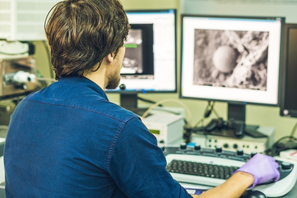
Electron microscopy is one of the main characterization methods used in nanotechnology research, development and manufacturing due to its excellent spatial resolution. In this article, AZoNano discusses liquid phase electron microscopy.

Image Credit: Elizaveta Galitckaia/Shutterstock.com
Electron Microscopy in Nanotechnology
The ability to achieve spatial resolutions that are greater than is possible in standard optical microscopy is what makes electron microscopy so important for the investigation of nanoscale structures but in terms of mapping their spatial and elemental configuration.1
A typical electron microscopy experiment is performed with the sample under high vacuum conditions. The use of a high vacuum serves several purposes. One, it prevents arcing of the electron guns using high voltages to provide the energetic electron beams for the experiment.
A high vacuum also means surfaces remain uncoated by contaminants in the environment and the electrons that are transmitted or scattered do not undergo secondary collisions before being detected.2
Given the power of electron microscopy for solid samples and their characterization, there has been interest in extending this to liquid phase samples, in particular, with interest in thin-film and microchip technologies.3
As electron microscopy images can be acquired as a function of time, one application of electron microscopy is to perform a time-resolved measurement to follow processes such as nanoparticle growth in real-time from the solution phase and other starting materials.4
Advancements in sample handling and the ability to perform electron microscopy under lower vacuum conditions have opened up a wealth of possibilities in terms of performing liquid phase electron microscopy measurements, including watching chemical reactions as they occur in solution.5
Liquid Phase Electron Microscopy
There are two main approaches to performing liquid phase electron microscopy measurements. Which method is suitable for the experiment depends largely on the volatility of the samples to be measured. Involatile liquid samples, like ionic liquids, do not evaporate readily, even under vacuum conditions. As a result, samples can be measured on standard electron microscopy instruments with no major modifications.
For volatile samples, the situation is more complicated. If the sample is volatile but not excessively so, environmental scanning electron microscopy may be appropriate. While the electron beam is produced in a similar way to a standard electron microscopy experiment, an environmental scanning microscopy experiment has additional vacuum considerations to deal with the high pressures in the sample region.
Environmental scanning electron microscopes typically have a series of differential pumping to cope with the high vacuum conditions needed in certain regions of the instrument and high local pressures near the outgassing samples. It is also necessary to consider some elements of the detector design to ensure the detector type is compatible with the generally higher electron counts and more complex pressure and chemical conditions in the environmental scanning electron microscopy instrument.
Sample Holders in Liquid Phase Electron Microscopy
Another approach for highly volatile samples that cannot be exposed to pressures lower than ambient or for measuring phenomena that occur at ambient conditions is to use specially designed sample cells for measurements. These samples cells can include a thin graphene shell that contributes little to the electron signal but protects the sample from the vacuum environment or completely sealed cells that have windows to allow the electron beam to pass through.5
More sophisticated cell designs can either be used to statically hold a liquid in the electron beam or for flow measurements, where liquids are either refreshed to compensate for electron damage or to do mixtures of reactants.
For nanoscience applications, electron microscopy is often used as a tool to look at the aggregation or the formation of agglomerates, even as the process occurs. With many electron microscopy sample cells, it is also possible to deliberately introduce moisture into the cell to demonstrate how real ambient conditions affect the sample behavior or what effects the presence of water has on the substrate at an atomic level.
The Future of Liquid Phase Electron Microscopy
There are now many commercially available solutions for the sample delivery for liquid phase electron microscopy, making it increasingly straightforward to make liquid phase measurements even on standard electron microscopy instruments.
One possibility is that with more complex graphene cell designs for electron microscopy, nanoscience applications can study micro or nanofluidics processes.6 Microfluidics instruments are now one approach to performing more specific and efficient chemical reactions, ideally with greater levels of reaction control achievable with more standard bulk chemical reactors.
Another very active area of research in liquid phase electron microscopy is to develop microscopy methodologies that can be used to look at nanofabrication processes. The idea would be that time-resolved electron microscopy could be used to watch structures on this length scale being formed, and this information can be used to provide active feedback on manufacturing processes.
Electron microscopy instrumentation remains a significant expense, with the cheapest instrument costing tens of thousands of pounds. There are now substantial efforts to find ways to make the instrumentation more affordable, particularly following the interest in cryo-electron microscopy as a tool for looking at biological structures.
Most other structurally sensitive experimental methods that can achieve similar resolution require access to advanced light sources, making electron microscopy an attractive option for laboratory or factory-based measurements.
References and Further Reading
Wang, Z. L. (2003). New Developments in Transmission Electron Microscopy for Nanotechnology. Advanced Materials, 18, 1497–1514. https://doi.org/10.1002/adma.200300384
Hawkes, P. W., & Spence, J. C. (Eds.). (2019). Springer handbook of microscopy (pp. 1064-1071). Cham, Switzerland: Springer International Publishing. https://doi.org/10.1007/978-3-030-00069-1
Bharda, A. V., & Jung, H. S. (2019). Liquid electron microscopy : then , now and future. Applied Microscopy, 49, 9. https://doi.org/10.1186/s42649-019-0011-7
Liao, H., Niu, K., & Zheng, H. (2013). Observation of growth of metal nanoparticles. Chemical Communications, 49, 11720. https://doi.org/10.1039/c3cc47473a
Kashin, A. S., & Ananikov, V. P. (2019). Monitoring chemical reactions in liquid media using electron microscopy. Nature Reviews Chemistry, 3, 624–637. https://doi.org/10.1038/s41570-019-0133-z
Dunn, G., Adiga, V. P., Pham, T., Bryant, C., Horton-bailey, D. J., Belling, J. N., Lafrance, B., Jackson, J. A., Barzegar, H. R., Yuk, J. M., Aloni, S., Crommie, M. F., & Zettl, A. (2020). Graphene-Sealed Flow Cells for In Situ Transmission Electron Microscopy of Liquid Samples. ACS Nano, 14, 9637–9643. https://doi.org/10.1021/acsnano.0c00431
Disclaimer: The views expressed here are those of the author expressed in their private capacity and do not necessarily represent the views of AZoM.com Limited T/A AZoNetwork the owner and operator of this website. This disclaimer forms part of the Terms and conditions of use of this website.