Jan 31 2011
Researchers from the University of Alabama at Birmingham's School of Engineering have created a three-dimensional electrospun scaffold on the nano scale that more effectively and efficiently facilitates cell and tissue growth in the laboratory.
Nanoscaffolds support the adhesion, growth and function of various cell types as they mature into specific tissues such as tendons, muscles and bones during tissue engineering. Yet, the traditional industry method for electrospinning creates densely packed sheet-like structures that prevent cells from penetrating the nanoscaffolds.
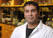
 Ajay Tambralli.
"Our three-dimensional electrospun nanoscaffolds better mimic nature and encourage cells to live longer and generate more viable, or functional, tissues," says Bryan Adam Blakeney, a recent graduate of the UAB Department of Biomedical Engineering and a lead author on the study recently published online by the journal Biomaterials.
The discovery could lead to new applications for the regeneration of a damaged pancreas due to diabetes, among other tissues and body parts damaged by disease or other causes.
To achieve its three-dimensional scaffold, which it calls a FLUF - for focused, low-density, uncompressed nanofibrous mesh - the research team uses a spherical dish with a slight curvature similar to that of a home TV satellite during the electrospinning process.
"This allows the nanofibers that constitute the scaffold to intertwine and accumulate without becoming too tightly packed, which is the primary problem with flat, two-dimensional scaffolds," says another lead author Ajay Tambralli, a student in the UAB School of Medicine.
Blakeney and Tambralli, who were both UAB biomedical engineering undergraduates when they conducted the research, and their faculty mentor Ho-Wook Jun, Ph.D., assistant professor of biomedical engineering, worked two years to refine the process.
The researchers say the FLUF's more loosely packed nanofibers encourage cells to grow through the scaffold structure, creating the more stable formations that better mimic natural tissue.
"Think of it like a bowl of rocks: You pour a liquid into the bowl, and the liquid fills all the large gaps between the rocks," Blakeney said. "But with our 3-D nanoscaffold, our rocks, or the fibers that constitute the scaffold, are the size of sand particles. We've separated the grains of sand to create gaps, and the cells fill and penetrate the gaps between the sand grains to form tissues."
In addition to Blakeney, Tambralli and Jun, co-authors on the study include biomedical engineering graduate student trainees Joel Anderson and Adinarayana Andukuri; biomedical engineering graduate research assistant Dong-Jin Lim; and Derrick Dean, Ph.D., an associate professor in the UAB Department of Materials Science and Engineering.
Source: http://www.uab.edu/